|
||
| 12. Digestive System | ||
| 1 2 3 4 5 6 7 8 9 10 11 12 13 14 15 16 17 18 19 20 21 22 23 24 25 | ||
| 26 27 28 29 30 31 32 33 34 35 36 37 38 39 40 41 42 43 44 45 46 47 48 49 50 | ||
| 51 52 53 54 55 56 57 58 59 60 61 62 63 64 65 66 67 68 69 70 71 72 73 74 75 | ||
| 76 77 78 79 80 81 82 83 84 85 86 |
| |||
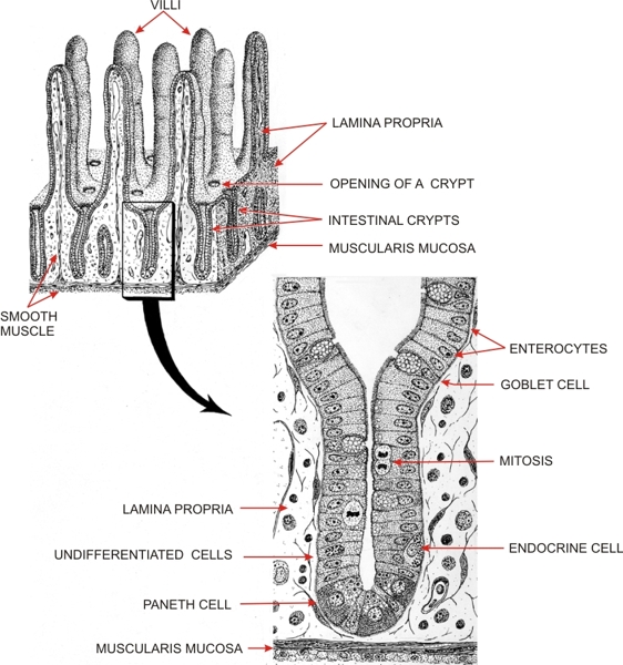 |
Diagram of the mucosa of the small intestine showing a three-dimensional representation of the mucosa (top left) and the components of an intestinal crypt (of Lieberkühn) (bottom right).
The upper part of the mucosa shows digitlike intestinal villi. The deeper part of the mucosa is occupied by tubular glands or crypts which open at the base of the villi. The mucosa is delimited by the muscularis mucosa. The intestinal crypt is lined with a simple columnar epithelium. At its base are zymogenic Paneth cells (which secrete bacteriolytic enzymes) and endocrine cells. Other cells of the crypt are undifferentiated. They proliferate and undergo renewal. Some of the daughter cells transform into goblet cells which migrate toward the mouth of the crypt, while others differentiate into absorptive cells or enterocytes which also migrate toward the villi. Other cells migrate toward the base of the crypt where they give rise to Paneth cells. Enterocytes and goblet cells migrate along the wall of the villi up to their tips where they are extruded. The renewal of the whole intestinal epithelium lasts from three to four days in rodents and five to six days in humans. N.B.: The proliferation, renewal and differentiation of the epithelial cells of the small intestine have continually been clarified by Dr. C. P. Leblond and his collaborators since 1950.
|
||