Contents Yves Clermont, Michael Lalli, Zsuzsanna Bencsath-Makkai
LIGHT-MICROSCOPIC HISTOLOGY ATLAS
     12. Digestive System
  1     2     3     4     5     6     7     8     9     10     11     12     13     14     15     16     17     18     19     20     21     22     23     24     25  
  26     27     28     29     30     31     32     33     34     35     36     37     38     39     40     41     42     43     44     45     46     47     48     49     50  
  51     52     53     54     55     56     57     58     59     60     61     62     63     64     65     66     67     68     69     70     71     72     73     74     75  
  76     77     78     79     80     81     82     83     84     85     86  

    12.33   Small intestine Version française
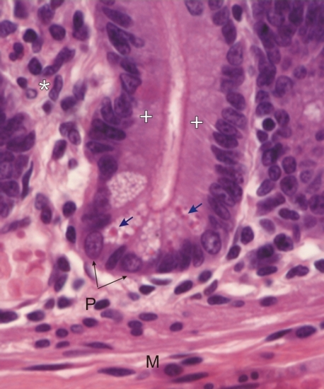
Section of the intestinal mucosa.

This field shows the bottom of an intestinal gland with a few Paneth cells (P) (bottom) and undifferentiated epithelial cells (+) (top).

Paneth cells show a basophilic cytoplasm around the nucleus and above a vacuolated area showing a few acidophilic secretory granules (arrows). The other granules were extracted during the histological procedure.

The lamina propria (*) and the muscularis mucosa (M) are also labelled.

Stain: H–­­E
Magnification: ×1100


We'd very much appreciate receiving your comments and suggestions!We'd very much appreciate receiving your comments and suggestions!

Creative Commons Licence The text and images of this Histology Atlas, by Yves Clermont, Michael Lalli & Zsuzsanna Bencsath-Makkai, are licensed under a Creative Commons Attribution-Noncommercial-No Derivative Works 2.5 Canada Licence and cannot be modified without the written permission of the authors. Use of any text or images must carry an acknowledgement which includes a link to the original work.


HAweb/Zs B-M -> HAbase-generated on 2013-09-13