Contents Yves Clermont, Michael Lalli, Zsuzsanna Bencsath-Makkai
LIGHT-MICROSCOPIC HISTOLOGY ATLAS
     12. Digestive System
  1     2     3     4     5     6     7     8     9     10     11     12     13     14     15     16     17     18     19     20     21     22     23     24     25  
  26     27     28     29     30     31     32     33     34     35     36     37     38     39     40     41     42     43     44     45     46     47     48     49     50  
  51     52     53     54     55     56     57     58     59     60     61     62     63     64     65     66     67     68     69     70     71     72     73     74     75  
  76     77     78     79     80     81     82     83     84     85     86  

    12.47   Mouse colon Version française
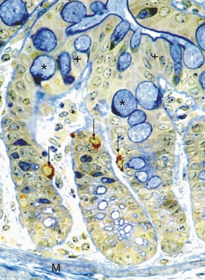
Mouse colon stained with toluidine blue and silver.

The bottoms of the crypts are seen next to the muscularis mucosa (M) below. The lumen of the colon is at the top of the image.

Among the differentiating epithelial cells of the crypts are endocrine cells showing reddish-brown secretory granules (arrows).

At the tops of the crypts, the goblet cells are fully differentiated and their granules are stained blue (*).

Between the goblet cells (*) the fully differentiated enterocytes (+) show their striated borders intensely stained blue-green.

Stain: Silver, toluidine blue
Magnification: ×850


We'd very much appreciate receiving your comments and suggestions!We'd very much appreciate receiving your comments and suggestions!

Creative Commons Licence The text and images of this Histology Atlas, by Yves Clermont, Michael Lalli & Zsuzsanna Bencsath-Makkai, are licensed under a Creative Commons Attribution-Noncommercial-No Derivative Works 2.5 Canada Licence and cannot be modified without the written permission of the authors. Use of any text or images must carry an acknowledgement which includes a link to the original work.


HAweb/Zs B-M -> HAbase-generated on 2013-09-13