|
||
| 12. Digestive System | ||
| 1 2 3 4 5 6 7 8 9 10 11 12 13 14 15 16 17 18 19 20 21 22 23 24 25 | ||
| 26 27 28 29 30 31 32 33 34 35 36 37 38 39 40 41 42 43 44 45 46 47 48 49 50 | ||
| 51 52 53 54 55 56 57 58 59 60 61 62 63 64 65 66 67 68 69 70 71 72 73 74 75 | ||
| 76 77 78 79 80 81 82 83 84 85 86 |
| |||
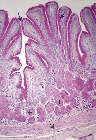 |
Dog pylorus stained with PAS.
The mucin of the surface mucous cells (arrows) that line the deep pits of the mucosa are stained dark violet-red in this field. The epithelial cells of the glands (+) are also positive PAS-positive, but their staining is less intense than that of the surface mucous cells lining the pits. The muscularis mucosa (M) delimiting the mucosa from the submucosa is lighly stained with PAS. Stain: PAS-Hematoxylin
|
||