Contents Yves Clermont, Michael Lalli, Zsuzsanna Bencsath-Makkai
LIGHT-MICROSCOPIC HISTOLOGY ATLAS
     12. Digestive System
  1     2     3     4     5     6     7     8     9     10     11     12     13     14     15     16     17     18     19     20     21     22     23     24     25  
  26     27     28     29     30     31     32     33     34     35     36     37     38     39     40     41     42     43     44     45     46     47     48     49     50  
  51     52     53     54     55     56     57     58     59     60     61     62     63     64     65     66     67     68     69     70     71     72     73     74     75  
  76     77     78     79     80     81     82     83     84     85     86  
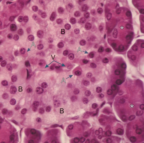
    12.63   Pancreas Version française
Pancreas of a dog.

Most of this field is occupied by a pancreatic islet of Langerhans.

In this section stained with hematoxylin and eosin, two types of cells can be identified: type B (B) and type A (A) cells. Type B cells are the more abundant and show a pale vaguely granulated cytoplasm. These cells secrete insulin. Type A cells, which are less abundant, have a cytoplasm containing fine acidophilic granules. They secrete glucagon.

Other cells, such as type D cells, which secrete somatostatin, can be identified only by using immunostaining methods.

A few collapsed capillaries (V) are present in the islet and glandular acini (*) surround the islet.

Stain: H–­­E
Magnification: ×1000


We'd very much appreciate receiving your comments and suggestions!We'd very much appreciate receiving your comments and suggestions!

Creative Commons Licence The text and images of this Histology Atlas, by Yves Clermont, Michael Lalli & Zsuzsanna Bencsath-Makkai, are licensed under a Creative Commons Attribution-Noncommercial-No Derivative Works 2.5 Canada Licence and cannot be modified without the written permission of the authors. Use of any text or images must carry an acknowledgement which includes a link to the original work.


HAweb/Zs B-M -> HAbase-generated on 2013-09-13