Contents Yves Clermont, Michael Lalli, Zsuzsanna Bencsath-Makkai
LIGHT-MICROSCOPIC HISTOLOGY ATLAS
     12. Digestive System
  1     2     3     4     5     6     7     8     9     10     11     12     13     14     15     16     17     18     19     20     21     22     23     24     25  
  26     27     28     29     30     31     32     33     34     35     36     37     38     39     40     41     42     43     44     45     46     47     48     49     50  
  51     52     53     54     55     56     57     58     59     60     61     62     63     64     65     66     67     68     69     70     71     72     73     74     75  
  76     77     78     79     80     81     82     83     84     85     86  

    12.40   Small intestine Version française
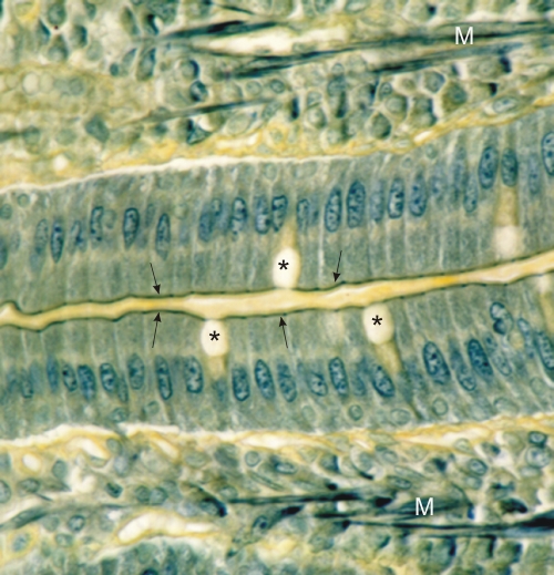
Section of a rodent intestinal mucosa stained with TPA.

This field shows the epithelial lining of two adjacent villi. The columnar enterocytes show their TPA-positive elongated nuclei and, at their apex, the TPA-positive terminal web. This intra-cytoplasmic web (arrows) is composed of fine cytoskeletal filaments that insert on the junctional terminal bars. In this preparation the striated borders are unstained and not visible. The lateral cell walls are equally pale.

The apical pockets of mucigen granules (*) of goblet cells are unstained.

The TPA-positive threads in the lamina propria correspond to smooth muscle fibres (M).

Stain: TPA
Magnification: ×950


We'd very much appreciate receiving your comments and suggestions!We'd very much appreciate receiving your comments and suggestions!

Creative Commons Licence The text and images of this Histology Atlas, by Yves Clermont, Michael Lalli & Zsuzsanna Bencsath-Makkai, are licensed under a Creative Commons Attribution-Noncommercial-No Derivative Works 2.5 Canada Licence and cannot be modified without the written permission of the authors. Use of any text or images must carry an acknowledgement which includes a link to the original work.


HAweb/Zs B-M -> HAbase-generated on 2013-09-13