Contents Yves Clermont, Michael Lalli, Zsuzsanna Bencsath-Makkai
LIGHT-MICROSCOPIC HISTOLOGY ATLAS
     12. Digestive System
  1     2     3     4     5     6     7     8     9     10     11     12     13     14     15     16     17     18     19     20     21     22     23     24     25  
  26     27     28     29     30     31     32     33     34     35     36     37     38     39     40     41     42     43     44     45     46     47     48     49     50  
  51     52     53     54     55     56     57     58     59     60     61     62     63     64     65     66     67     68     69     70     71     72     73     74     75  
  76     77     78     79     80     81     82     83     84     85     86  

    12.21   Stomach Version française
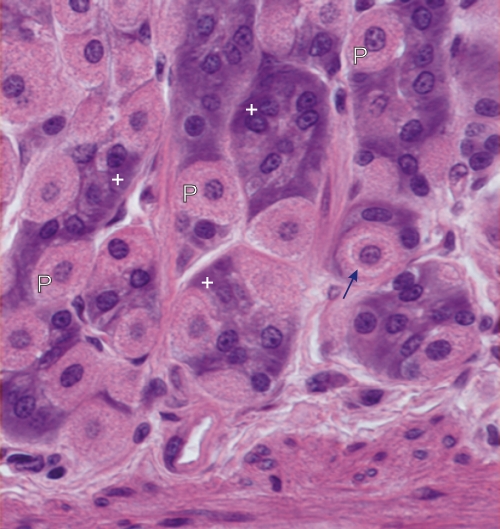
Gastric mucosa of a dog.

At the base of the gastric glands the zymogenic cells (+) are deeply stained with hematoxylin and the parietal cells (P) are well stained with eosin, due to their acidophilic mitochondria.

In one of the well-stained parietal cells, a lightly stained perinuclear intracellular canaliculus (arrow) is clearly visible. This small canaliculus, which opens at the apex of the cell, is the site of the transmembrane flux of ions that leads to the formation of HCl in the lumen of the gland.

Stain: H–­­E
Magnification: ×1100


We'd very much appreciate receiving your comments and suggestions!We'd very much appreciate receiving your comments and suggestions!

Creative Commons Licence The text and images of this Histology Atlas, by Yves Clermont, Michael Lalli & Zsuzsanna Bencsath-Makkai, are licensed under a Creative Commons Attribution-Noncommercial-No Derivative Works 2.5 Canada Licence and cannot be modified without the written permission of the authors. Use of any text or images must carry an acknowledgement which includes a link to the original work.


HAweb/Zs B-M -> HAbase-generated on 2013-09-13