Contents Yves Clermont, Michael Lalli, Zsuzsanna Bencsath-Makkai
LIGHT-MICROSCOPIC HISTOLOGY ATLAS
     12. Digestive System
  1     2     3     4     5     6     7     8     9     10     11     12     13     14     15     16     17     18     19     20     21     22     23     24     25  
  26     27     28     29     30     31     32     33     34     35     36     37     38     39     40     41     42     43     44     45     46     47     48     49     50  
  51     52     53     54     55     56     57     58     59     60     61     62     63     64     65     66     67     68     69     70     71     72     73     74     75  
  76     77     78     79     80     81     82     83     84     85     86  

    12.81   Bile canaliculi Version française
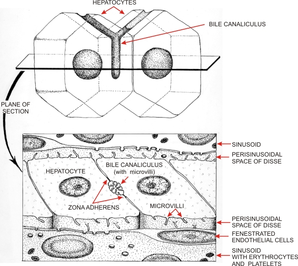
Diagrams illustrating the localization and structure of a bile canaliculus at the interface of two hepatocytes. The bottom drawing represents the bile canaliculus as seen with the electron microscope.

The bile canaliculus forms hemicylindrical grooves facing adjacent hepatocytes. It is delimited by zona or zonula adherens which tightly bind the cytoplasmic membranes of facing hepatocytes. The lumen of the canaliculus shows numerous microvilli.

Facing the blood sinusoids, lined with fenestrated endothelial cells, the cytoplasmic membrane of the hepatocytes shows short microvilli which occupy the narrow perisinusoidal space of Disse (see Figure 12.84.


We'd very much appreciate receiving your comments and suggestions!We'd very much appreciate receiving your comments and suggestions!

Creative Commons Licence The text and images of this Histology Atlas, by Yves Clermont, Michael Lalli & Zsuzsanna Bencsath-Makkai, are licensed under a Creative Commons Attribution-Noncommercial-No Derivative Works 2.5 Canada Licence and cannot be modified without the written permission of the authors. Use of any text or images must carry an acknowledgement which includes a link to the original work.


HAweb/Zs B-M -> HAbase-generated on 2013-09-13