|
||
12. Digestive System | ||
1 2 3 4 5 6 7 8 9 10 11 12 13 14 15 16 17 18 19 20 21 22 23 24 25 | ||
26 27 28 29 30 31 32 33 34 35 36 37 38 39 40 41 42 43 44 45 46 47 48 49 50 | ||
51 52 53 54 55 56 57 58 59 60 61 62 63 64 65 66 67 68 69 70 71 72 73 74 75 | ||
76 77 78 79 80 81 82 83 84 85 86 |
| |||
![]() |
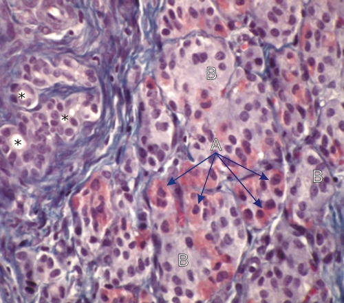
Section of a dog pancreas.
This animal had its main pancreatic duct ligatured for several weeks. In this experimental condition the digestive enzymes released by the degenerating zymogenic cells led to a complete lysis of the acini. The dispersed islets of Langerhans, which resist lysis, merged into an irregular mass. In this islet tissue, acidophilic A cells (A) can be identified among the pale B cells (B). Some residual clustered ducts are also visible (*). Stain: Masson’s Trichrome
|
The text and images of this Histology Atlas, by Yves Clermont,
Michael Lalli & Zsuzsanna Bencsath-Makkai,
are licensed under a
Creative Commons Attribution-Noncommercial-No Derivative Works 2.5 Canada Licence
and cannot be modified without the written permission of the authors.
Use of any text or images must carry an acknowledgement which includes a link to the original work.