Contents Yves Clermont, Michael Lalli, Zsuzsanna Bencsath-Makkai
LIGHT-MICROSCOPIC HISTOLOGY ATLAS
     12. Digestive System
  1     2     3     4     5     6     7     8     9     10     11     12     13     14     15     16     17     18     19     20     21     22     23     24     25  
  26     27     28     29     30     31     32     33     34     35     36     37     38     39     40     41     42     43     44     45     46     47     48     49     50  
  51     52     53     54     55     56     57     58     59     60     61     62     63     64     65     66     67     68     69     70     71     72     73     74     75  
  76     77     78     79     80     81     82     83     84     85     86  

    12.5   Esophageal-cardiac junction Version française
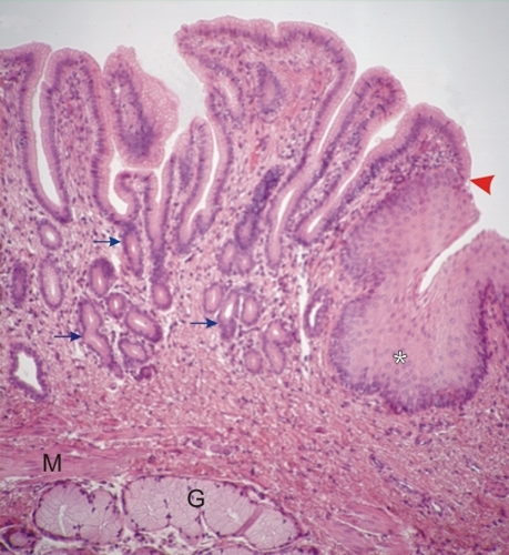
Junction of the esophagus to the cardiac stomach in humans.

The stratified squamous epithelium of the esophagus (*) changes abruptly (arrowhead) to the simple columnar epithelium (left) lining the cardiac portion of the gastric mucosa.

This mucosa shows deep recesses or pits continuous with the tubular cardiac glands (arrows).

The muscularis mucosa (M), composed of smooth muscle fibres, separates the mucosa from the submucosa. The submucosa contains esophageal mucous glands (G).

Stain: H–­­E
Magnification: ×250


We'd very much appreciate receiving your comments and suggestions!We'd very much appreciate receiving your comments and suggestions!

Creative Commons Licence The text and images of this Histology Atlas, by Yves Clermont, Michael Lalli & Zsuzsanna Bencsath-Makkai, are licensed under a Creative Commons Attribution-Noncommercial-No Derivative Works 2.5 Canada Licence and cannot be modified without the written permission of the authors. Use of any text or images must carry an acknowledgement which includes a link to the original work.


HAweb/Zs B-M -> HAbase-generated on 2013-09-13