|
||
| 12. Digestive System | ||
| 1 2 3 4 5 6 7 8 9 10 11 12 13 14 15 16 17 18 19 20 21 22 23 24 25 | ||
| 26 27 28 29 30 31 32 33 34 35 36 37 38 39 40 41 42 43 44 45 46 47 48 49 50 | ||
| 51 52 53 54 55 56 57 58 59 60 61 62 63 64 65 66 67 68 69 70 71 72 73 74 75 | ||
| 76 77 78 79 80 81 82 83 84 85 86 |
| |||
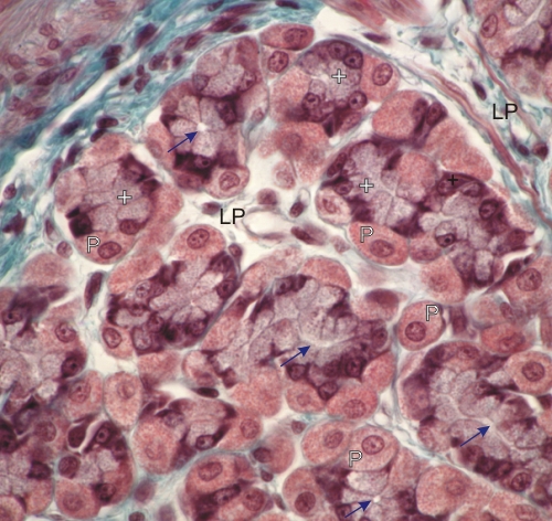 |
Gastric mucosa of a dog.
This field shows transverse or oblique sections through the base of several gastric glands. Zymogenic cells (+) and acidophilic parietal cells (P) are visible. The narrow central excretory ducts of the glands are also visible (arrows). These glands are surrounded by the connective tissue of the lamina propria (LP). Stain: Massons Trichrome
|
||