|
||
| 12. Digestive System | ||
| 1 2 3 4 5 6 7 8 9 10 11 12 13 14 15 16 17 18 19 20 21 22 23 24 25 | ||
| 26 27 28 29 30 31 32 33 34 35 36 37 38 39 40 41 42 43 44 45 46 47 48 49 50 | ||
| 51 52 53 54 55 56 57 58 59 60 61 62 63 64 65 66 67 68 69 70 71 72 73 74 75 | ||
| 76 77 78 79 80 81 82 83 84 85 86 |
| |||
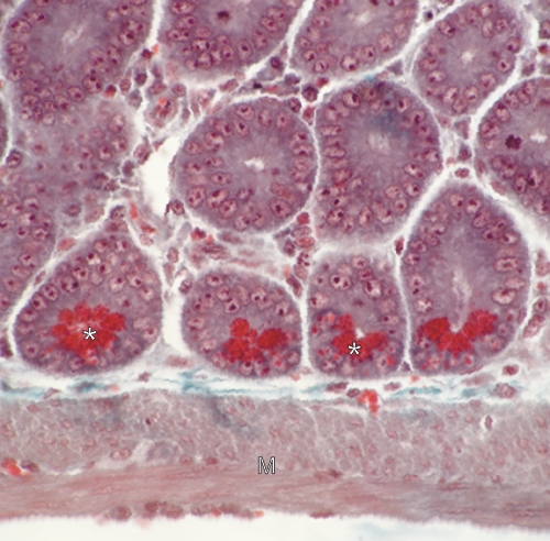 |
Section of the intestinal mucosa of a bat.
In this section the intestinal crypts are cut transversely. The bottoms of the crypts are next to the muscularis mucosa (M). The Paneth cells (*) at the very bottom of the crypts show their well-preserved acidophilic secretory granules. The muscularis mucosa shows two layers of muscle cells, the inner circular one is seen in cross section and the outer longitudinal one is seen in a longitudinal section. Stain: Massons Trichrome
|
||