Contents Yves Clermont, Michael Lalli, Zsuzsanna Bencsath-Makkai
LIGHT-MICROSCOPIC HISTOLOGY ATLAS
     12. Digestive System
  1     2     3     4     5     6     7     8     9     10     11     12     13     14     15     16     17     18     19     20     21     22     23     24     25  
  26     27     28     29     30     31     32     33     34     35     36     37     38     39     40     41     42     43     44     45     46     47     48     49     50  
  51     52     53     54     55     56     57     58     59     60     61     62     63     64     65     66     67     68     69     70     71     72     73     74     75  
  76     77     78     79     80     81     82     83     84     85     86  

    12.13   Stomach Version française
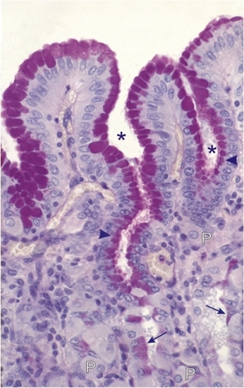
Gastric mucosa of a monkey.

This field shows gastric pits (*) lined with surface mucous cells containing an increasing amount of PAS-positive mucigen granules from the bottom of the pits to the surface of the mucosa (arrowheads).

A few PAS-positive mucous neck cells (arrows) of gastric glands are also seen. Some lightly stained parietal cells (P) are also present along the wall of the glands.

Stain: PAS-Hematoxylin
Magnification: ×1000


We'd very much appreciate receiving your comments and suggestions!We'd very much appreciate receiving your comments and suggestions!

Creative Commons Licence The text and images of this Histology Atlas, by Yves Clermont, Michael Lalli & Zsuzsanna Bencsath-Makkai, are licensed under a Creative Commons Attribution-Noncommercial-No Derivative Works 2.5 Canada Licence and cannot be modified without the written permission of the authors. Use of any text or images must carry an acknowledgement which includes a link to the original work.


HAweb/Zs B-M -> HAbase-generated on 2013-09-13