|
||
| 12. Digestive System | ||
| 1 2 3 4 5 6 7 8 9 10 11 12 13 14 15 16 17 18 19 20 21 22 23 24 25 | ||
| 26 27 28 29 30 31 32 33 34 35 36 37 38 39 40 41 42 43 44 45 46 47 48 49 50 | ||
| 51 52 53 54 55 56 57 58 59 60 61 62 63 64 65 66 67 68 69 70 71 72 73 74 75 | ||
| 76 77 78 79 80 81 82 83 84 85 86 |
| |||
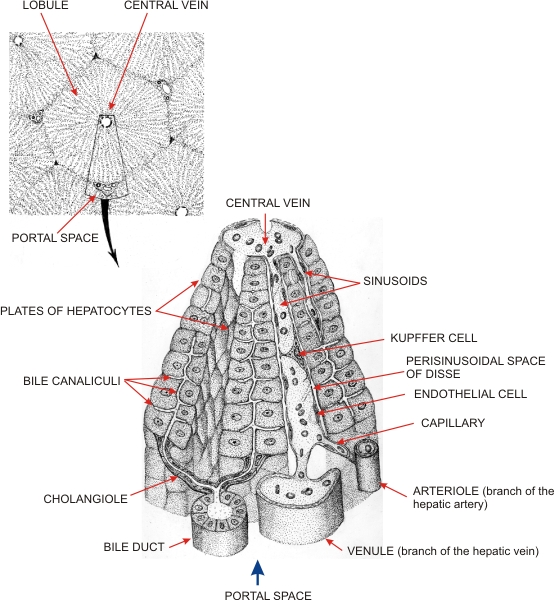 |
Schematic representations of the liver to illustrate the relations of hepatocyte plates to bile ducts and to blood vessels (i.e., portal veins, hepatic arteries, sinusoids and central veins).
The small diagram shows, at low power, the outline of a lobule with the central vein in the centre and, in some corners of the hexagonal lobule, portal spaces occupied by branches of the portal veins, hepatic arteries and bile ducts. The larger diagram, which corresponds to the framed area of the small diagram, illustrates hepatocyte plates (left) that extend from the portal space to the central vein. Some of these plates are schematically represented as double rows of hepatocytes to demonstrate the system of intercellular bile canaliculi. These canaliculi are located at the interfaces of hepatocytes in the centre of the single plates of cells. (Left) The networks of canaliculi connect with narrow ducts, called cholangioles, which are lined with squamous epithelial cells. These ducts are equivalent to the intercalated ducts of the pancreas. They open into bile ducts located in the portal spaces. These portal bile ducts are lined with a simple cuboidal epithelium. (Right) Branches of portal veins and hepatic arteries open into the sinusoids, which are lined with fenestrated endothelial cells and large macrophages called Kupffer cells. The blood flows in these sinusoidal meanders and reaches the central vein at the top.
|
||