|
||
| 12. Digestive System | ||
| 1 2 3 4 5 6 7 8 9 10 11 12 13 14 15 16 17 18 19 20 21 22 23 24 25 | ||
| 26 27 28 29 30 31 32 33 34 35 36 37 38 39 40 41 42 43 44 45 46 47 48 49 50 | ||
| 51 52 53 54 55 56 57 58 59 60 61 62 63 64 65 66 67 68 69 70 71 72 73 74 75 | ||
| 76 77 78 79 80 81 82 83 84 85 86 |
| |||
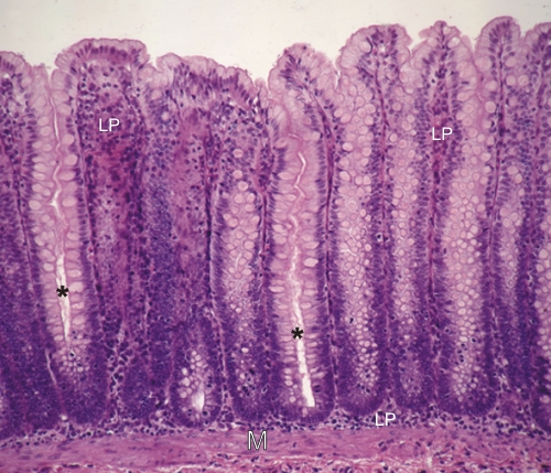 |
Mucosa of a dog colon.
The intestinal crypts are cut longitudinally but only two present a lumen (*). The others show their epithelium cut tangentially. The goblet cells and enterocytes migrate from the base of the crypt to the surface of the mucosa where they are extruded. The lamina propria (LP) and the muscularis mucosa (M) are also labelled. Stain: HE
|
||