Contents Yves Clermont, Michael Lalli, Zsuzsanna Bencsath-Makkai
LIGHT-MICROSCOPIC HISTOLOGY ATLAS
     12. Digestive System
  1     2     3     4     5     6     7     8     9     10     11     12     13     14     15     16     17     18     19     20     21     22     23     24     25  
  26     27     28     29     30     31     32     33     34     35     36     37     38     39     40     41     42     43     44     45     46     47     48     49     50  
  51     52     53     54     55     56     57     58     59     60     61     62     63     64     65     66     67     68     69     70     71     72     73     74     75  
  76     77     78     79     80     81     82     83     84     85     86  

    12.9   Stomach Version française
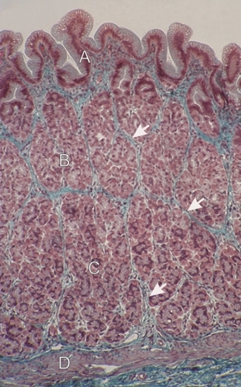
Oblique section of the gastric mucosa of a dog.

The layer of pits (A) shows mucosal folds lined with surface mucous cells sitting on the lamina propria.

The gastric glands show the necks (B) and bases (C) cut obliquely.

Groups of glands are delimited by the connective tissue septae, stained green, of the lamina propria (arrows).

The muscularis mucosa (D) is composed of smooth muscle fibres.

Stain: Masson’s Trichrome
Magnification: ×130


We'd very much appreciate receiving your comments and suggestions!We'd very much appreciate receiving your comments and suggestions!

Creative Commons Licence The text and images of this Histology Atlas, by Yves Clermont, Michael Lalli & Zsuzsanna Bencsath-Makkai, are licensed under a Creative Commons Attribution-Noncommercial-No Derivative Works 2.5 Canada Licence and cannot be modified without the written permission of the authors. Use of any text or images must carry an acknowledgement which includes a link to the original work.


HAweb/Zs B-M -> HAbase-generated on 2013-09-13