Contents Yves Clermont, Michael Lalli, Zsuzsanna Bencsath-Makkai
LIGHT-MICROSCOPIC HISTOLOGY ATLAS
     12. Digestive System
  1     2     3     4     5     6     7     8     9     10     11     12     13     14     15     16     17     18     19     20     21     22     23     24     25  
  26     27     28     29     30     31     32     33     34     35     36     37     38     39     40     41     42     43     44     45     46     47     48     49     50  
  51     52     53     54     55     56     57     58     59     60     61     62     63     64     65     66     67     68     69     70     71     72     73     74     75  
  76     77     78     79     80     81     82     83     84     85     86  

    12.28   Duodenum Version française
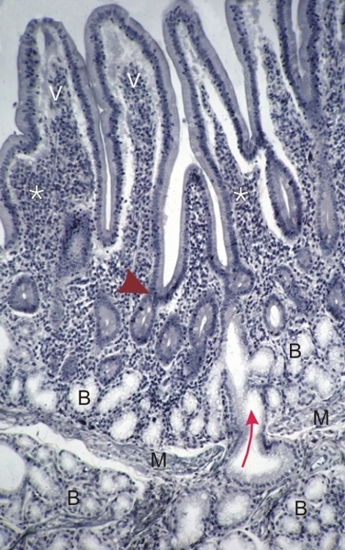
Section of the duodenum.

The muscularis mucosa (M) is seen at the borderline of the mucosa above and the submucosa below.

The mucosa shows the intestinal villi (V) and the intestinal crypts, which open at their base (arrowhead).

Mucous acini of Brunner's glands (B) are seen in both the mucosa and the submucosa. Some cross the muscularis mucosa (arrow) and open into an intestinal crypt.

The lamina propria (*) of the mucosa contains many lymphoid cells.

Stain: Iron Hematoxylin
Magnification: ×270


We'd very much appreciate receiving your comments and suggestions!We'd very much appreciate receiving your comments and suggestions!

Creative Commons Licence The text and images of this Histology Atlas, by Yves Clermont, Michael Lalli & Zsuzsanna Bencsath-Makkai, are licensed under a Creative Commons Attribution-Noncommercial-No Derivative Works 2.5 Canada Licence and cannot be modified without the written permission of the authors. Use of any text or images must carry an acknowledgement which includes a link to the original work.


HAweb/Zs B-M -> HAbase-generated on 2013-09-13