|
||
| 12. Digestive System | ||
| 1 2 3 4 5 6 7 8 9 10 11 12 13 14 15 16 17 18 19 20 21 22 23 24 25 | ||
| 26 27 28 29 30 31 32 33 34 35 36 37 38 39 40 41 42 43 44 45 46 47 48 49 50 | ||
| 51 52 53 54 55 56 57 58 59 60 61 62 63 64 65 66 67 68 69 70 71 72 73 74 75 | ||
| 76 77 78 79 80 81 82 83 84 85 86 |
| |||
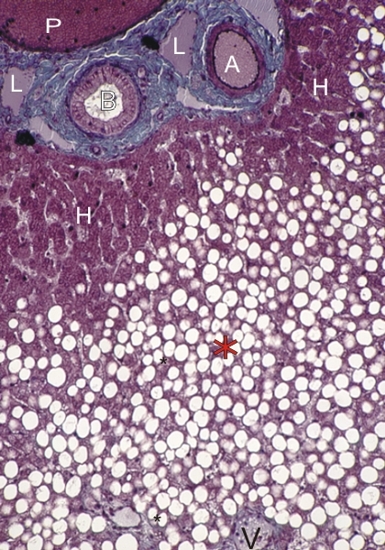 |
Section of the liver of a dog.
This animal was administered a toxic substance that provokes the accumulation of fat in hepatocytes. This treatment emphasizes the zonation of the liver lobule, owing to the selective damage to some hepatocytes produced by the toxic substance administered. While normal plates of chromophilic hepatocytes (H) are seen next to a portal space, the fatty degeneration of hepatocytes (*) is observed around the central vein (V) of the lobule. The following structures in the connective tissue (blue-green) of the portal space are labelled:
Stain: Massons Trichrome
|
||