|
||
| 6. Lymphatic Organs | ||
| 1 2 3 4 5 6 7 8 9 10 11 12 13 14 15 16 17 18 19 20 21 22 23 24 25 | ||
| 26 27 28 29 30 31 32 33 34 35 36 37 38 |
| |||
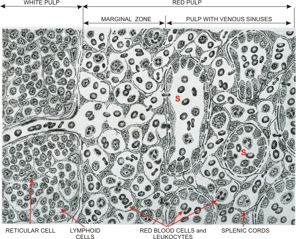 |
Drawing illustrating the composition of the spleen with the white pulp (left) and the red pulp (right).
The red pulp shows the marginal zone free of venous sinuses and the remaining red pulp with numerous venous sinuses (S). The white pulp is a dense lymphatic tissue with lymphoid cells located in spaces between reticular cells and fibres. It includes capillaries that open directly into the marginal zone. This marginal zone is composed of small reticular cells and fine reticular fibres infiltrated with blood cells, platelets and macrophages. The blood cells percolate through this marginal zone and reach the red pulp containing venous sinuses. These sinuses are separated by the splenic cords (of Billroth). The blood cells which do not degenerate during their travel through the red pulp return to the blood circulation through gaps between the endothelial cells of the sinuses (see Figure 5.32C). The spleen thus serves to filter the blood by eliminating senescent blood cells and platelets.
|
||