Contents Yves Clermont, Michael Lalli, Zsuzsanna Bencsath-Makkai
LIGHT-MICROSCOPIC HISTOLOGY ATLAS
     6. Lymphatic Organs
  1     2     3     4     5     6     7     8     9     10     11     12     13     14     15     16     17     18     19     20     21     22     23     24     25  
  26     27     28     29     30     31     32     33     34     35     36     37     38  

    6.35   Spleen Version française
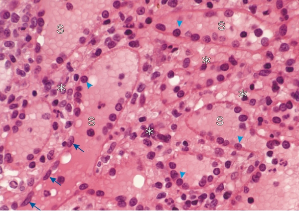
Section of the red pulp of the spleen.

This field shows the part of the red pulp containing numerous venous sinuses (S). These vessels are identified either by their endothelial cells showing elongated ovoid nuclei (arrows) in a longitudinal section of the sinus or as circular profiles (arrowheads) in cross or oblique sections of the sinus.

These venous sinuses are separated by the splenic cords (*) composed of reticular cells and fibres, macrophages and various circulating blood elements.

Stain: H–­­E
Magnification: ×600


We'd very much appreciate receiving your comments and suggestions!We'd very much appreciate receiving your comments and suggestions!

Creative Commons Licence The text and images of this Histology Atlas, by Yves Clermont, Michael Lalli & Zsuzsanna Bencsath-Makkai, are licensed under a Creative Commons Attribution-Noncommercial-No Derivative Works 2.5 Canada Licence and cannot be modified without the written permission of the authors. Use of any text or images must carry an acknowledgement which includes a link to the original work.


HAweb/Zs B-M -> HAbase-generated on 2013-09-13