|
||
| 6. Lymphatic Organs | ||
| 1 2 3 4 5 6 7 8 9 10 11 12 13 14 15 16 17 18 19 20 21 22 23 24 25 | ||
| 26 27 28 29 30 31 32 33 34 35 36 37 38 |
| |||
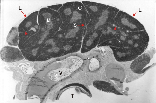 |
Transverse section of a rat thymus stained with Dominicis method.
In proximity to the trachea (T) and a large vessel (V), the thymus shows two main lobes separated by a thin continuous septum (S). Each lobe is subdivided into lobules (L) by incomplete septa (arrowheads) connected to the capsule covering the organ. The tissue of the thymus shows a deeply stained cortex (C) and a pale medulla (M). Stain: Toluidine blue, eosin
|
||