Contents Yves Clermont, Michael Lalli, Zsuzsanna Bencsath-Makkai
LIGHT-MICROSCOPIC HISTOLOGY ATLAS
     6. Lymphatic Organs
  1     2     3     4     5     6     7     8     9     10     11     12     13     14     15     16     17     18     19     20     21     22     23     24     25  
  26     27     28     29     30     31     32     33     34     35     36     37     38  

    6.24   Lymph nodule Version française
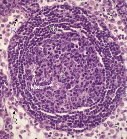
Section of a lymphatic nodule.

This field shows a lymphatic nodule with a germinal centre (G) surrounded by the mantle (Ma).

This nodule located in the deep cortex is surrounded here by a lymphatic sinus (S).

The germinal centre of the nodule shows the following cell types: large, medium and small lymphocytes and a few macrophages (Mac). The mantle is composed of concentric layers of small lymphocytes. In this field the limit between the mantle and the surrounding diffuse lymphatic tissue is not evident.

Reticular cells (R) are labelled in the sinuses.

Stain: H–­­E
Magnification: ×450


We'd very much appreciate receiving your comments and suggestions!We'd very much appreciate receiving your comments and suggestions!

Creative Commons Licence The text and images of this Histology Atlas, by Yves Clermont, Michael Lalli & Zsuzsanna Bencsath-Makkai, are licensed under a Creative Commons Attribution-Noncommercial-No Derivative Works 2.5 Canada Licence and cannot be modified without the written permission of the authors. Use of any text or images must carry an acknowledgement which includes a link to the original work.


HAweb/Zs B-M -> HAbase-generated on 2013-09-13