|
||
| 6. Lymphatic Organs | ||
| 1 2 3 4 5 6 7 8 9 10 11 12 13 14 15 16 17 18 19 20 21 22 23 24 25 | ||
| 26 27 28 29 30 31 32 33 34 35 36 37 38 |
| |||
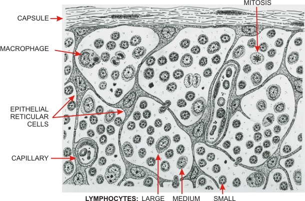 |
Drawing illustrating the composition of thymic cortex.
The connective tissue capsule is shown at the top. The framework of the cortex is composed of stellate cells attached to each other by desmosomes at the tips of their processes (not visible with the light microscope). These cells, which are of epithelial origin, contain filaments of keratin similar to those in other epithelial cells and are called epithelial reticular cells. The spaces between these stellate cells are occupied by lymphocytes of various sizes. Large lymphocytes, which are rare, multiply to give rise to medium lymphocytes which in turn divide several times to yield numerous small thymic lymphocytes, or T-lymphocytes. These lymphocytes migrate from the cortex to the medulla of the thymus. Macrophages are also present in the cortex and contribute to the elimination by phagocytosis of the residues of degenerating lymphocytes.
|
||