Contents Yves Clermont, Michael Lalli, Zsuzsanna Bencsath-Makkai
LIGHT-MICROSCOPIC HISTOLOGY ATLAS
     6. Lymphatic Organs
  1     2     3     4     5     6     7     8     9     10     11     12     13     14     15     16     17     18     19     20     21     22     23     24     25  
  26     27     28     29     30     31     32     33     34     35     36     37     38  

    6.3   Thymus Version française
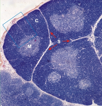
Section of a rat thymus stained with Dominici’s method.

The connective tissue septa (S) demarcate three lobules, each showing an intensely stained cortex (C) and a lightly stained medulla (M).

Although the medulla appears discontinuous and isolated by the cortex in this section, the medulla is in fact continuous through the lobe as revealed by the examination of serial sections.

The framed area is shown at a higher magnification in Figure 6.4.

Stain: Toluidine blue, eosin
Magnification: ×80


We'd very much appreciate receiving your comments and suggestions!We'd very much appreciate receiving your comments and suggestions!

Creative Commons Licence The text and images of this Histology Atlas, by Yves Clermont, Michael Lalli & Zsuzsanna Bencsath-Makkai, are licensed under a Creative Commons Attribution-Noncommercial-No Derivative Works 2.5 Canada Licence and cannot be modified without the written permission of the authors. Use of any text or images must carry an acknowledgement which includes a link to the original work.


HAweb/Zs B-M -> HAbase-generated on 2013-09-13