Contents Yves Clermont, Michael Lalli, Zsuzsanna Bencsath-Makkai
LIGHT-MICROSCOPIC HISTOLOGY ATLAS
     6. Lymphatic Organs
  1     2     3     4     5     6     7     8     9     10     11     12     13     14     15     16     17     18     19     20     21     22     23     24     25  
  26     27     28     29     30     31     32     33     34     35     36     37     38  

    6.28   Lymph node medulla Version française
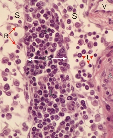
Section of the medulla of a lymph node.

In this section, the medullary cord (Co) is almost completely depleted of plasma cells and a large majority of the lymphoid cells are lymphocytes (L). The cellular composition of the cord thus indicates that this node is minimally reactive to antigens.

The sinuses (S) contain reticular cells (R) and lymphocytes (L). In the sinus on the right, an arteriole (A) and a venule (V) are seen in connective tissue trabecules.

Stain: H–­­E
Magnification: ×700


We'd very much appreciate receiving your comments and suggestions!We'd very much appreciate receiving your comments and suggestions!

Creative Commons Licence The text and images of this Histology Atlas, by Yves Clermont, Michael Lalli & Zsuzsanna Bencsath-Makkai, are licensed under a Creative Commons Attribution-Noncommercial-No Derivative Works 2.5 Canada Licence and cannot be modified without the written permission of the authors. Use of any text or images must carry an acknowledgement which includes a link to the original work.


HAweb/Zs B-M -> HAbase-generated on 2013-09-13