Contents Yves Clermont, Michael Lalli, Zsuzsanna Bencsath-Makkai
LIGHT-MICROSCOPIC HISTOLOGY ATLAS
     6. Lymphatic Organs
  1     2     3     4     5     6     7     8     9     10     11     12     13     14     15     16     17     18     19     20     21     22     23     24     25  
  26     27     28     29     30     31     32     33     34     35     36     37     38  

    6.38   Spleen Version française
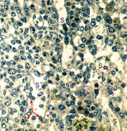
Section of the red pulp of a rodent spleen stained with TPA.

This method stains green the nuclei of cells and, in the cytoplasm of the endothelial cells (arrowheads) of venous sinuses (S), their basal plates. These basal plates are cytoplasmic cytoskeletal elements that demarcate the basal limits of these endothelial cells.

These cells have spaces between them (see also Figure 5.32C) through which leukocytes and red blood cells migrate to return to the blood circulation (triple arrow).

The splenic cords are labelled with asterisks.

Stain: TPA
Magnification: ×650


We'd very much appreciate receiving your comments and suggestions!We'd very much appreciate receiving your comments and suggestions!

Creative Commons Licence The text and images of this Histology Atlas, by Yves Clermont, Michael Lalli & Zsuzsanna Bencsath-Makkai, are licensed under a Creative Commons Attribution-Noncommercial-No Derivative Works 2.5 Canada Licence and cannot be modified without the written permission of the authors. Use of any text or images must carry an acknowledgement which includes a link to the original work.


HAweb/Zs B-M -> HAbase-generated on 2013-09-13