|
||
| 5. Vessels | ||
| 1 2 3 4 5 6 7 8 9 10 11 12 13 14 15 16 17 18 19 20 21 22 23 24 25 | ||
| 26 27 28 29 30 31 32 33 34 35 |
| |||
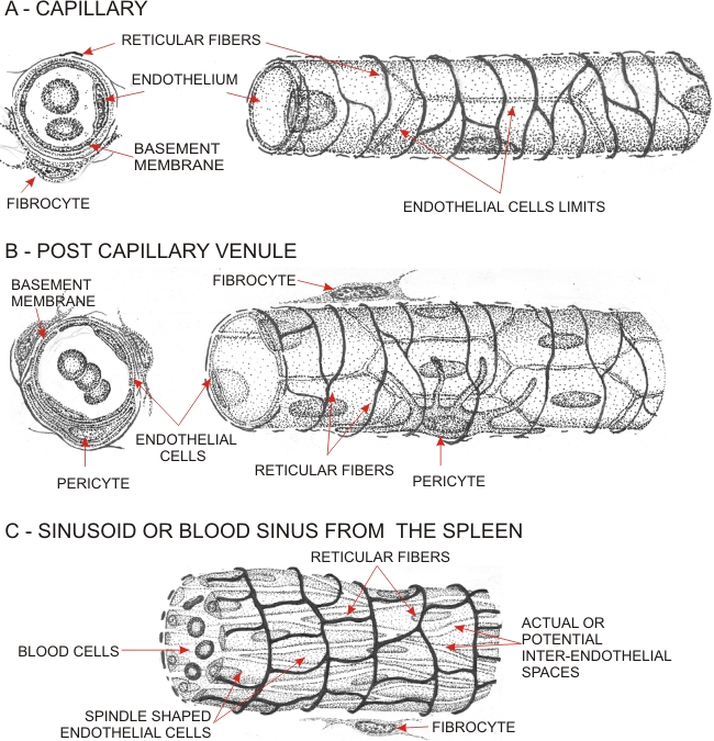 |
Drawings illustrating the structure of three small vessels:
The two vessels above are shown in cross sections (left) and in three dimensions (right). The argyrophilic reticular fibres (type III collagen) are included. The basement membranes associated with the endothelial cells of the capillary and post-capillary venule are indicated in the cross sections. A basement membrane is absent from the wall of sinusoids. The capillaries are composed of endothelial cells with their edges closely apposed to each other. The post-capillary venules leave actual or potential gaps at the interfaces of endothelial cells thus creating possible passageways for the migration of leukocytes. The post-capillary venules are often associated with stellate contractile cells, or pericytes, which are enclosed by a basement membrane. The sinusoids (or blood sinuses) have large irregular luminae. In the spleen, as illustrated here, the endothelial cells are not squamous but are spindle-shaped and hemi-cylindrical. There are potential gaps between them that permit the passage of white and red blood cells. These endothelial cells are not associated with a basement membrane but are held in place by a large network of thick reticular fibres.
|
||