|
||
| 6. Lymphatic Organs | ||
| 1 2 3 4 5 6 7 8 9 10 11 12 13 14 15 16 17 18 19 20 21 22 23 24 25 | ||
| 26 27 28 29 30 31 32 33 34 35 36 37 38 |
| |||
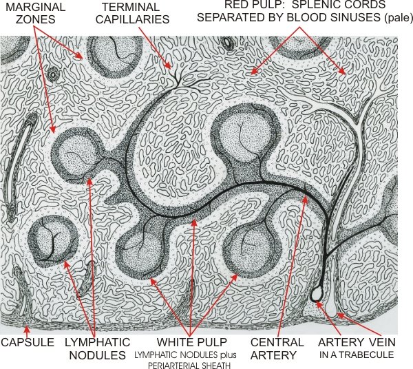 |
Drawing of a small portion of a spleen to illustrate the two major compartments of the organ: the white pulp and the red pulp.
The spleen is covered with a connective tissue capsule that extends inside the organ as trabecules. Some of these trabecules serve as passageways for arteries and veins. From there, arteries enter the white pulp of the spleen and branch into arterioles and capillaries. The white pulp is divided into two areas: the periarterial lymphatic sheath and the lymphatic nodules. The latter, similar to the nodules of the lymph nodes, show a germinal centre and a mantle or corona. The red pulp shows two areas: the marginal zone, which is free of venous sinuses and is next to the white pulp; and the remaining red pulp showing numerous venous sinuses separated by the splenic cords. Some of the terminal capillaries of the white pulp open directly into the venous sinuses or terminate in the splenic cords. Other capillaries end at the periphery of the white pulp and release the blood into the marginal zone. Blood cells eventually reach the sinuses of the red pulp. The venous sinuses are continuous with venules which connect to the veins returning to the surface of the spleen via the trabecules. There is no lymphatic vessel inside the spleen.
|
||