Contents Yves Clermont, Michael Lalli, Zsuzsanna Bencsath-Makkai
LIGHT-MICROSCOPIC HISTOLOGY ATLAS
     6. Lymphatic Organs
  1     2     3     4     5     6     7     8     9     10     11     12     13     14     15     16     17     18     19     20     21     22     23     24     25  
  26     27     28     29     30     31     32     33     34     35     36     37     38  

    6.4   Thymus Version française
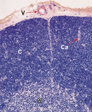
Section of a rat thymus stained with Dominici’s method. This field is a higher magnification of the framed area in Figure 6.3.

At this magnification it is evident that the heavy staining of the cortex (C) is due to the closely packed small lymphocytes with their basophilic cytoplasm and nucleus.

The medulla (M) also contains lymphocytes, but these cells are spaced by larger cells that are lightly stained.

A vessel (V) in the capsule and capillaries (Ca) crossing the cortex are also identified.

Stain: Toluidine blue, eosin
Magnification: ×400


We'd very much appreciate receiving your comments and suggestions!We'd very much appreciate receiving your comments and suggestions!

Creative Commons Licence The text and images of this Histology Atlas, by Yves Clermont, Michael Lalli & Zsuzsanna Bencsath-Makkai, are licensed under a Creative Commons Attribution-Noncommercial-No Derivative Works 2.5 Canada Licence and cannot be modified without the written permission of the authors. Use of any text or images must carry an acknowledgement which includes a link to the original work.


HAweb/Zs B-M -> HAbase-generated on 2013-09-13