Contents Yves Clermont, Michael Lalli, Zsuzsanna Bencsath-Makkai
LIGHT-MICROSCOPIC HISTOLOGY ATLAS
     6. Lymphatic Organs
  1     2     3     4     5     6     7     8     9     10     11     12     13     14     15     16     17     18     19     20     21     22     23     24     25  
  26     27     28     29     30     31     32     33     34     35     36     37     38  

    6.29   Lymph node medulla Version française
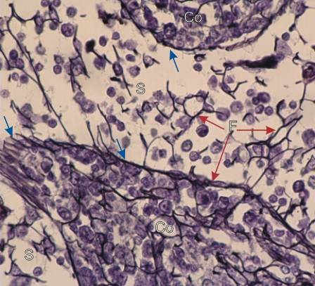
Section of the medulla of a lymph node stained with silver to show reticular fibres (type III collagen) in black.

Note that the reticular fibres (F) are present in both the sinuses (S) and the medullary cords (Co). There is an accumulation of reticular fibres along the edges of the cord (blue arrows).

Nuclei of reticular cells are closely related to the reticular fibres.

Stain: Hematoxylin and silver
Magnification: ×700


We'd very much appreciate receiving your comments and suggestions!We'd very much appreciate receiving your comments and suggestions!

Creative Commons Licence The text and images of this Histology Atlas, by Yves Clermont, Michael Lalli & Zsuzsanna Bencsath-Makkai, are licensed under a Creative Commons Attribution-Noncommercial-No Derivative Works 2.5 Canada Licence and cannot be modified without the written permission of the authors. Use of any text or images must carry an acknowledgement which includes a link to the original work.


HAweb/Zs B-M -> HAbase-generated on 2013-09-13