Contents Yves Clermont, Michael Lalli, Zsuzsanna Bencsath-Makkai
LIGHT-MICROSCOPIC HISTOLOGY ATLAS
     6. Lymphatic Organs
  1     2     3     4     5     6     7     8     9     10     11     12     13     14     15     16     17     18     19     20     21     22     23     24     25  
  26     27     28     29     30     31     32     33     34     35     36     37     38  

    6.36   Spleen Version française
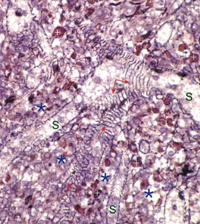
Section of a rodent spleen stained with silver to show the reticular fibres in black.

In the red pulp the venous sinuses (S) can be identified by the thick reticular fibres forming supporting networks (arrows) around the sinuses.

The brownish cells in this section are macrophages (+) located in the splenic cords (*). These cells are pigmented in brown by hemosiderin, which is a breakdown product of hemoglobin derived from lysed red blood cells.

Stain: Hematoxylin and silver
Magnification: ×700


We'd very much appreciate receiving your comments and suggestions!We'd very much appreciate receiving your comments and suggestions!

Creative Commons Licence The text and images of this Histology Atlas, by Yves Clermont, Michael Lalli & Zsuzsanna Bencsath-Makkai, are licensed under a Creative Commons Attribution-Noncommercial-No Derivative Works 2.5 Canada Licence and cannot be modified without the written permission of the authors. Use of any text or images must carry an acknowledgement which includes a link to the original work.


HAweb/Zs B-M -> HAbase-generated on 2013-09-13