|
||
| 6. Lymphatic Organs | ||
| 1 2 3 4 5 6 7 8 9 10 11 12 13 14 15 16 17 18 19 20 21 22 23 24 25 | ||
| 26 27 28 29 30 31 32 33 34 35 36 37 38 |
| |||
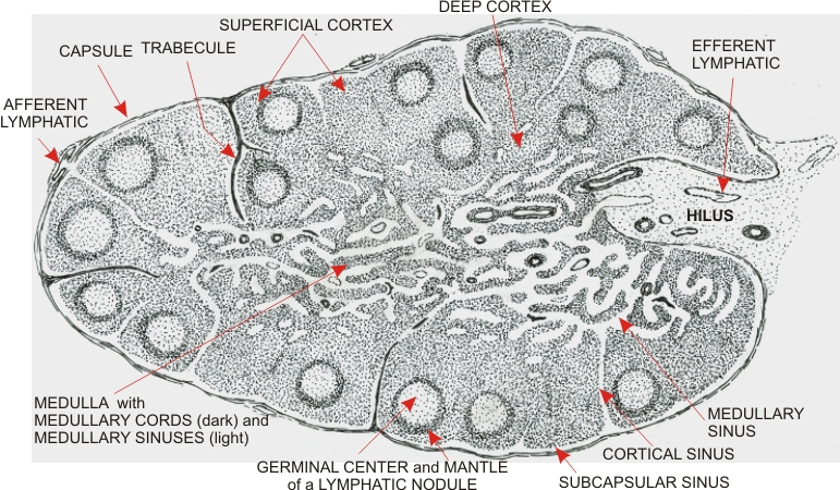 |
Drawing of a lymph node based on the section shown in Figure 6.14.
This organ is delimited by a connective tissue capsule which extends inside the node as trabecules. Afferent lymphatic vessels located in the capsule open into a system of interconnected lymphatic sinuses that traverse the organ and exit from its hilus to enter the efferent lymphatics. The lymph node is subdivided into two areas: the cortex and the medulla. The cortex is composed of lymphatic nodules showing a germinal centre and a denser mantle. Between and below the nodules, the lymphatic tissue is more diffuse and occupies the rest of the cortex. The medulla is composed of cordlike extensions of the diffuse lymphatic tissue of the cortex. These anastomotic medullary cords are spaced by medullary sinuses that direct the lymph toward the hilus.
|
||