|
||
| 6. Lymphatic Organs | ||
| 1 2 3 4 5 6 7 8 9 10 11 12 13 14 15 16 17 18 19 20 21 22 23 24 25 | ||
| 26 27 28 29 30 31 32 33 34 35 36 37 38 |
| |||
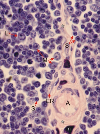 |
Section of a rat thymus stained with Dominici's method.
This field shows a connective tissue septum with some cortex on each side. This cortex includes some large lymphocytes (L), medium lymphocytes (M) and small lymphocytes (S). The nuclei and cytoplasm of epithelial reticular cells are masked by lymphocytes but some of them (ER) are seen applied to the connective tissue septum. An arteriole (A) and a mast cell (m) are labelled in the septum. Stain: Toluidine blue, eosin
|
||