|
||
| 6. Lymphatic Organs | ||
| 1 2 3 4 5 6 7 8 9 10 11 12 13 14 15 16 17 18 19 20 21 22 23 24 25 | ||
| 26 27 28 29 30 31 32 33 34 35 36 37 38 |
| |||
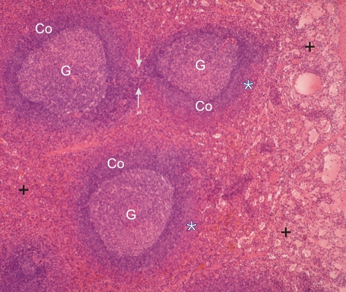 |
Section of the spleen.
In this field the white pulp appears as three nodules with their respective germinal centres (G) and mantles or coronas (Co). As illustrated in Figure 6.30, these nodules are connected by the white pulp (arrows). Around the white pulp, the red pulp shows strongly acidophilic marginal zones (*) free of venous sinuses. The rest of the red pulp shows numerous profiles of venous sinuses (+). Stain: HE
|
||