Contents Yves Clermont, Michael Lalli, Zsuzsanna Bencsath-Makkai
LIGHT-MICROSCOPIC HISTOLOGY ATLAS
     6. Lymphatic Organs
  1     2     3     4     5     6     7     8     9     10     11     12     13     14     15     16     17     18     19     20     21     22     23     24     25  
  26     27     28     29     30     31     32     33     34     35     36     37     38  

    6.12   Thymic medulla Version française
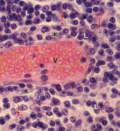
Section of the medulla of a human thymus.

This field shows a large venule (V) surrounded by a narrow perivascular connective tissue space (*).

This space is occupied by lymphocytes (L) which mask the fibrocyte-like reticular cells and fibres. The perivascular space serves as a passageway for lymphocytes which migrate from the epithelial compartment of the thymus to the lumen of the venule.

The nuclei of epithelial reticular cells (arrows) outside the perivascular space are identified. They are characterized by a large acidophilic nucleolus.

Stain: H–­­E
Magnification: ×1000


We'd very much appreciate receiving your comments and suggestions!We'd very much appreciate receiving your comments and suggestions!

Creative Commons Licence The text and images of this Histology Atlas, by Yves Clermont, Michael Lalli & Zsuzsanna Bencsath-Makkai, are licensed under a Creative Commons Attribution-Noncommercial-No Derivative Works 2.5 Canada Licence and cannot be modified without the written permission of the authors. Use of any text or images must carry an acknowledgement which includes a link to the original work.


HAweb/Zs B-M -> HAbase-generated on 2013-09-13