|
||
| 6. Lymphatic Organs | ||
| 1 2 3 4 5 6 7 8 9 10 11 12 13 14 15 16 17 18 19 20 21 22 23 24 25 | ||
| 26 27 28 29 30 31 32 33 34 35 36 37 38 |
| |||
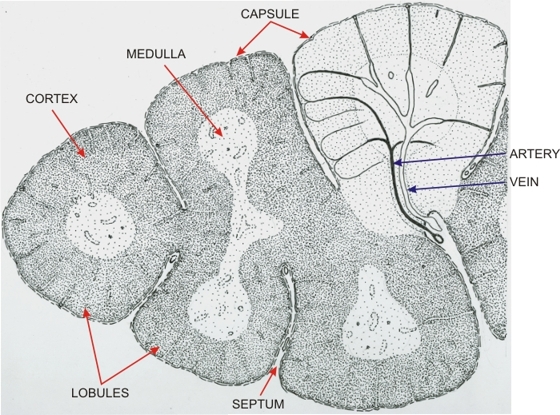 |
Drawing of a portion of a thymic lobe showing several lobules delimited by extensions of incomplete connective tissue septa continuous with the capsule of the organ.
The thymic tissue is subdivided into cortex and medulla. This drawing illustrates the blood circulation in the upper right lobule. An artery from the depth of a septum reaches the medulla by going through the cortex. In the medulla the arteries branch into arterioles that enter the cortex, subdivide and yield capillary loops that return to the medulla. These capillaries are continuous with venules which fuse to form veins that return to the septum. Other arterioles located in the capsule give rise to capillaries that cross the cortex and directly reach the medullary venules.
|
||