Contents Yves Clermont, Michael Lalli, Zsuzsanna Bencsath-Makkai
LIGHT-MICROSCOPIC HISTOLOGY ATLAS
     6. Lymphatic Organs
  1     2     3     4     5     6     7     8     9     10     11     12     13     14     15     16     17     18     19     20     21     22     23     24     25  
  26     27     28     29     30     31     32     33     34     35     36     37     38  

    6.25   Lymph node medulla Version française
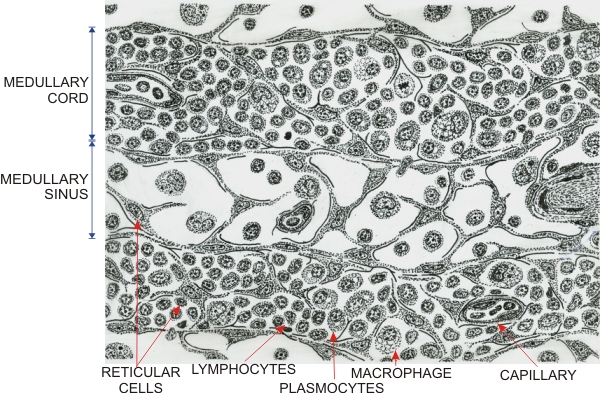
Drawing of the medulla of a lymph node showing two medullary cords separated by medullary lymphatic sinuses.

The composition of medullary sinuses is identical to that of cortical sinuses. They are composed of reticular cells and reticular fibres forming spaces occupied by circulating lymphocytes and macrophages.

In the medullary cords, reticular cells and fibres form a compartment occupied by large, medium and small lymphocytes and macrophages.

The medullary cords may also contain plasma cells secreting immunoglobulins. These cells derive from B-lymphocytes challenged by antigens. The number of plasma cells in lymph node cords depends on the degree of response to immunogenic antigens.


We'd very much appreciate receiving your comments and suggestions!We'd very much appreciate receiving your comments and suggestions!

Creative Commons Licence The text and images of this Histology Atlas, by Yves Clermont, Michael Lalli & Zsuzsanna Bencsath-Makkai, are licensed under a Creative Commons Attribution-Noncommercial-No Derivative Works 2.5 Canada Licence and cannot be modified without the written permission of the authors. Use of any text or images must carry an acknowledgement which includes a link to the original work.


HAweb/Zs B-M -> HAbase-generated on 2013-09-13