Contents Yves Clermont, Michael Lalli, Zsuzsanna Bencsath-Makkai
LIGHT-MICROSCOPIC HISTOLOGY ATLAS
     6. Lymphatic Organs
  1     2     3     4     5     6     7     8     9     10     11     12     13     14     15     16     17     18     19     20     21     22     23     24     25  
  26     27     28     29     30     31     32     33     34     35     36     37     38  

    6.27   Lymph node medulla Version française
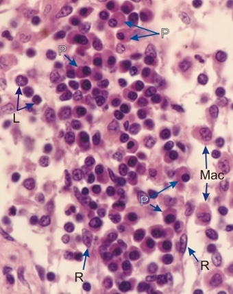
Section of the medulla of a lymph node.

The following cells are labelled in the medullary cord and sinuses: reticular cells (R), lymphocytes (L), plasma cells (P), macrophages (Mac).

The antibody producing plasma cells are ovoid cells with a small spherical eccentrically located nucleus in a chromophilic cytoplasm. The staining of the cytoplasm is due to an abundance of ribosomes associated with a profuse network of endoplasmic reticulum cisternae. A small spherical lightly stained juxtanuclear area containing some plasma cells (P) is occupied by the Golgi apparatus.

Stain: H–­­E
Magnification: ×800


We'd very much appreciate receiving your comments and suggestions!We'd very much appreciate receiving your comments and suggestions!

Creative Commons Licence The text and images of this Histology Atlas, by Yves Clermont, Michael Lalli & Zsuzsanna Bencsath-Makkai, are licensed under a Creative Commons Attribution-Noncommercial-No Derivative Works 2.5 Canada Licence and cannot be modified without the written permission of the authors. Use of any text or images must carry an acknowledgement which includes a link to the original work.


HAweb/Zs B-M -> HAbase-generated on 2013-09-13