Contents Yves Clermont, Michael Lalli, Zsuzsanna Bencsath-Makkai
LIGHT-MICROSCOPIC HISTOLOGY ATLAS
     6. Lymphatic Organs
  1     2     3     4     5     6     7     8     9     10     11     12     13     14     15     16     17     18     19     20     21     22     23     24     25  
  26     27     28     29     30     31     32     33     34     35     36     37     38  

    6.14   Lymph node Version française
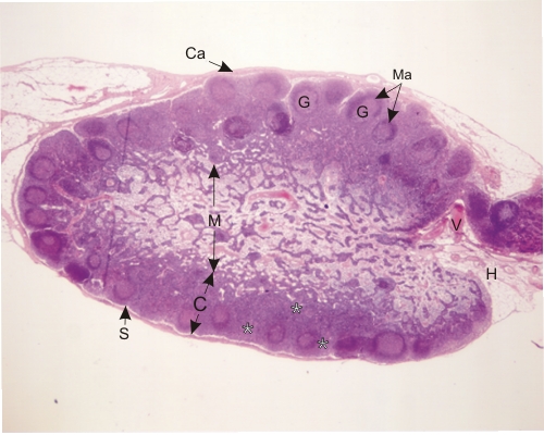
Section of a lymph node of a rodent.

The plane of section passes through the hilus (H). Several components of the node, as illustrated in the diagram of Figure 6.13, are labelled:

  • Capsule (Ca)
  • Cortex (C)
  • Subcapsular sinus (S)
  • Lymphatic nodules with their germinal centres (G) and mantle (Ma)
  • Diffuse cortical lymphatic tissue (*)
  • Medulla (M) with its medullary cords and sinuses
  • Vessels of the hilus (V)

Stain: H–­­E
Magnification: ×25


We'd very much appreciate receiving your comments and suggestions!We'd very much appreciate receiving your comments and suggestions!

Creative Commons Licence The text and images of this Histology Atlas, by Yves Clermont, Michael Lalli & Zsuzsanna Bencsath-Makkai, are licensed under a Creative Commons Attribution-Noncommercial-No Derivative Works 2.5 Canada Licence and cannot be modified without the written permission of the authors. Use of any text or images must carry an acknowledgement which includes a link to the original work.


HAweb/Zs B-M -> HAbase-generated on 2013-09-13