|
||
| 6. Lymphatic Organs | ||
| 1 2 3 4 5 6 7 8 9 10 11 12 13 14 15 16 17 18 19 20 21 22 23 24 25 | ||
| 26 27 28 29 30 31 32 33 34 35 36 37 38 |
| |||
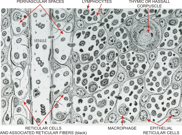 |
Drawing showing an area of the thymic medulla.
A post-capillary venule is surrounded by a perivascular connective tissue sheath on its left and by the epithelial compartment of the medulla on its right. The perivascular sheath is composed of fibrocyte-like reticular cells associated with reticular fibres and of lymphocytes and macrophages. The epithelial compartment includes epithelial reticular cells similar to those of the cortex. Some of them form concentric layers of spherical structures, called thymic corpuscles, or Hassalls corpuscles. These corpuscles are present only in the medulla of the thymus. Lymphocytes, some degenerating, and macrophages are present between the epithelial reticular cells. The T-lymphocytes, most of which originate in the cortex, migrate into the epithelial compartment of the medulla and cross the perivascular space to enter the lumen of the venule.
|
||