|
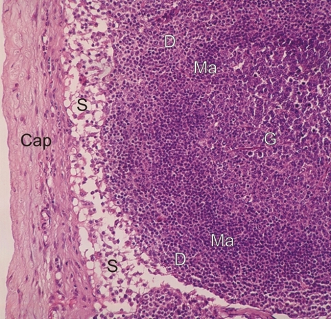
Section of the cortex of a lymph node stained with hematoxylin and eosin.
Underlying the connective tissue capsule (Cap), the lightly stained layer corresponds to a subcapsular sinus (S). Deeper, the dense lymphatic tissue shows a lymphatic nodule with a germinal centre (G) and a more intensely stained mantle (Ma). The lymphatic tissue around the mantle is the diffuse cortical lymphatic tissue (D).
Note that there are no sharp demarcations between the various zones of the cortical lymphatic tissue.
Stain: HE
Magnification: ×200
|