|
||
| 6. Lymphatic Organs | ||
| 1 2 3 4 5 6 7 8 9 10 11 12 13 14 15 16 17 18 19 20 21 22 23 24 25 | ||
| 26 27 28 29 30 31 32 33 34 35 36 37 38 |
| |||
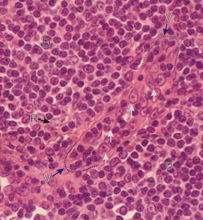 |
Section of the cortex of a lymph node.
This field shows a portion of the diffuse lymphatic tissue (D) which includes a longitudinal section of a post-capillary venule (V). The limits of the two extremities of this venule are indicated by broken lines. The endothelial cells (white arrows) of these vessels are thick rather than squamous as is typical for venules, so they are called high endothelial venules. These particular venules, seen exclusively in the diffuse lymphatic tissue of the lymph node cortex, are sites of migration of lymphocytes in and out of the blood circulation. The presence of small lymphocytes between the endothelial cells of these venules is the expression of such a migration. The elongated nucleus of a reticular cell (R) is labelled in the diffuse lymphatic tissue. Stain: HE
|
||