Sommaire Yves Clermont, Michael Lalli, Zsuzsanna Bencsath-Makkai
ATLAS D'HISTOLOGIE EN MICROSCOPIE OPTIQUE
     6. Organes Lymphatiques
  1     2     3     4     5     6     7     8     9     10     11     12     13     14     15     16     17     18     19     20     21     22     23     24     25  
  26     27     28     29     30     31     32     33     34     35     36     37     38  
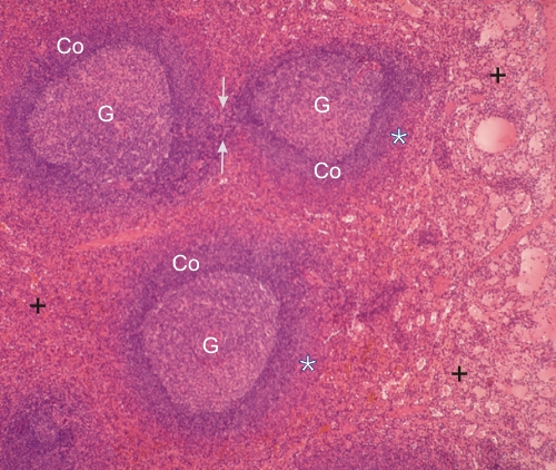
    6.31   Rate English version
Coupe de la rate.

Dans ce champ, la pulpe blanche montre trois nodules chacun avec un centre germinatif (G) et une couronne (Co) ou manteau.

Tel qu’indiqué dans la figure 6.30, ces nodules sont en continuité les uns avec les autres par du tissu lymphatique associé aux artères ou artérioles (flèches).

Les couches très acidophiles qui entourent les nodules correspondent aux zones marginales (*).

Le reste de la pulpe rouge contient d’innombrables sinus veineux (+).

Coloration: H–­­É
Grossissement: ×50


We'd very much appreciate receiving your comments and suggestions!We'd very much appreciate receiving your comments and suggestions!

Creative Commons Licence The text and images of this Histology Atlas, by Yves Clermont, Michael Lalli & Zsuzsanna Bencsath-Makkai, are licensed under a Creative Commons Attribution-Noncommercial-No Derivative Works 2.5 Canada Licence and cannot be modified without the written permission of the authors. Use of any text or images must carry an acknowledgement which includes a link to the original work.


HAweb/Zs B-M -> HAbase-generated on 2013-09-13