 |
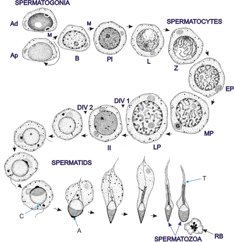
Spermatogenesis in man, that is, the formation of spermatozoa from spermatogonial stem cells, is divided into three stages:
- Proliferation, renewal and differentiation of spermatogonia taking place among the following cells:
- Dark type A spermatogonia (or reserve stem cells which give rise to type Ap when the needs arise) (Ad)
- Pale type A spermatogonia (or renewing stem cells) (Ap)
- Differentiated spermatogonia (B)
The spermatogonia Ap and B divide by mitosis (M). All type B cells give rise to preleptotene spermatocytes.
- Meiosis or reductional divisions involving primary and secondary spermatocytes. The prophase of the first meiotic division of primary spermatocytes go through the following steps:
preleptotene (Pl) → leptotene (L) → zygotene (Z) → early pachytene (EP) → mid pachytene (MP) → late pachytene (LP). The late pachtene cells undergo the first meiotic division that gives rise to secondary spermatocytes (II), which soon undergo the second meiotic divisions that yield haploid spermatids.
- Spermiogenesis, the metamorphosis of spermatids into spermatozoa.
The following structures are labelled: Golgi apparatus (G), acrosome (A), centrioles (C), tail (T) and residual body (RB).
Characteristics of human spermatogenesis: - In man the duration of spermatogenesis is fixed and lasts approximately 60 days.
- Stem cells enter spermatogenesis at regular intervals (every 16 days).
- Type Ap stem cells enter spermatogenesis in groups, the cells of such groups are connected by open intercellular bridges. Spermatocytes and spermatids which arise from such groups of spermatogonial stem cells are also connected by intercellular bridges and all the interconnected cells of a given generation (spermatocytes or spermatids) evolve synchronously.
- The epithelium of seminiferous tubules is composed of from four to five such generations of germinal cells at various steps of their evolution (see Figure 15.10).
|