|
||
| 15. Male Reproductive System | ||
| 1 2 3 4 5 6 7 8 9 10 11 12 13 14 15 16 17 18 19 20 21 22 23 24 25 | ||
| 26 27 28 29 30 31 32 33 34 35 36 37 38 39 40 41 42 43 44 |
| |||
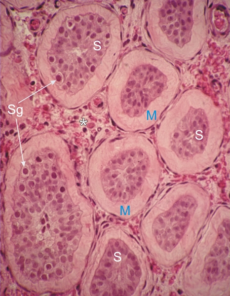 |
Section of the testis of a hypophysectomized adult human.
The atrophied seminiferous tubules show a thickened limiting membrane (M) composed of connective tissue fibres. The regressed epithelium that fills the rest of the tubule is composed mainly of regressed Sertoli cells (S) and a few spermatogonia (Sg). In the interstitial spaces (*) the endocrine Leydig cells are also atrophied in the absence of stimulation from the gonadotrophic hormones (compare with the Leydig cells in a normal human testis in Figure 15.16). Stain: HE
|
||