Contents Yves Clermont, Michael Lalli, Zsuzsanna Bencsath-Makkai
LIGHT-MICROSCOPIC HISTOLOGY ATLAS
     15. Male Reproductive System
  1     2     3     4     5     6     7     8     9     10     11     12     13     14     15     16     17     18     19     20     21     22     23     24     25  
  26     27     28     29     30     31     32     33     34     35     36     37     38     39     40     41     42     43     44  

    15.13   Human seminiferous tubule Version française
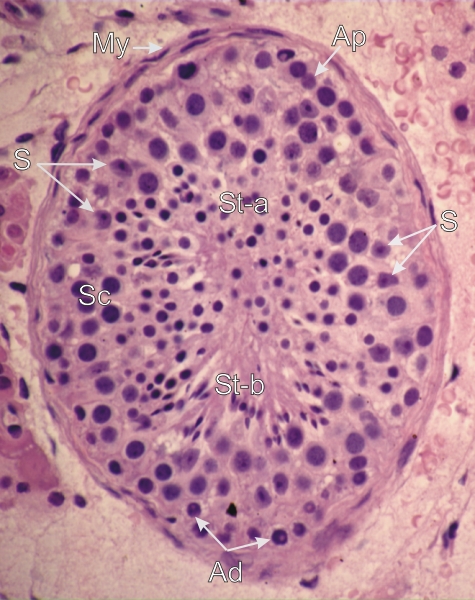
Section showing an overall view of a human seminiferous tubule and its limiting membrane (My).

Various generations of germinal cells are labelled:

  • Spermatogonia (Ad and Ap) are seen along the limiting membrane.
  • The nuclei of primary spermatocytes (Sc), which form a second layer, are mixed with nuclei of Sertoli cells (S).
  • Nuclei of early round spermatids (St-a) and of late elongated spermatids (St-b) are present toward the centre of the tubule and occlude the lumen.

The nuclei of myoid cells (My) are seen in the limiting membrane.

Stain: H–­­E
Magnification: ×600


We'd very much appreciate receiving your comments and suggestions!We'd very much appreciate receiving your comments and suggestions!

Creative Commons Licence The text and images of this Histology Atlas, by Yves Clermont, Michael Lalli & Zsuzsanna Bencsath-Makkai, are licensed under a Creative Commons Attribution-Noncommercial-No Derivative Works 2.5 Canada Licence and cannot be modified without the written permission of the authors. Use of any text or images must carry an acknowledgement which includes a link to the original work.


HAweb/Zs B-M -> HAbase-generated on 2013-09-13