|
||
| 15. Male Reproductive System | ||
| 1 2 3 4 5 6 7 8 9 10 11 12 13 14 15 16 17 18 19 20 21 22 23 24 25 | ||
| 26 27 28 29 30 31 32 33 34 35 36 37 38 39 40 41 42 43 44 |
| |||
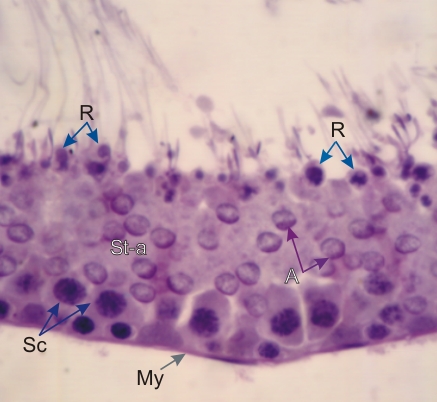 |
Rat seminiferous epithelium in a section stained with periodic acid- Schiff-hematoxylin.
PAS stains carbohydrates and emphasizes the glycoproteins contained in the acrosomes (A). Acrosomes, which cap the nuclei of spermatids (St-a), play an important role in the fertilization of the oocyte. Note the presence of basophilic residual bodies (R) along the surface of the epithelium. These cytoplasmic bodies separate from the spermatozoa, as these cells detach from the seminiferous epithelium (see Figure 15.10). These residual bodies are eliminated by the phagocytic activity of Sertoli cells. Some pachytene spermatocytes (Sc) are also labelled. Note that the thin limiting membrane of rat tubules is composed of a single layer of myoid cells (My). Stain: PAS-Hematoxylin
|
||