Contents Yves Clermont, Michael Lalli, Zsuzsanna Bencsath-Makkai
LIGHT-MICROSCOPIC HISTOLOGY ATLAS
     15. Male Reproductive System
  1     2     3     4     5     6     7     8     9     10     11     12     13     14     15     16     17     18     19     20     21     22     23     24     25  
  26     27     28     29     30     31     32     33     34     35     36     37     38     39     40     41     42     43     44  

    15.5   Seminiferous tubule Version française
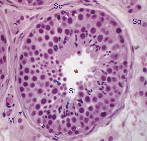
Cross section of a human seminiferous tubule.

Between the limiting membrane (M) and the tubular lumen (*) is a complex stratified epithelium.

This epithelium is composed of two main classes of cells: the supporting non-dividing somatic Sertoli cells (S) (see figures 15.10–15.13), and the germinal cells at various steps of spermatogenesis: spermatogonia (Sg), spermatocytes (Sc) and spermatids (St).

The limiting membrane (M) is composed of flattened contractile cells or myoid cells separated by a small amount of connective tissue fibrils (collagen, elastin, fibrilin and reticulin).

Stain: H–­­E
Magnification: ×400


We'd very much appreciate receiving your comments and suggestions!We'd very much appreciate receiving your comments and suggestions!

Creative Commons Licence The text and images of this Histology Atlas, by Yves Clermont, Michael Lalli & Zsuzsanna Bencsath-Makkai, are licensed under a Creative Commons Attribution-Noncommercial-No Derivative Works 2.5 Canada Licence and cannot be modified without the written permission of the authors. Use of any text or images must carry an acknowledgement which includes a link to the original work.


HAweb/Zs B-M -> HAbase-generated on 2013-09-13