Contents Yves Clermont, Michael Lalli, Zsuzsanna Bencsath-Makkai
LIGHT-MICROSCOPIC HISTOLOGY ATLAS
     15. Male Reproductive System
  1     2     3     4     5     6     7     8     9     10     11     12     13     14     15     16     17     18     19     20     21     22     23     24     25  
  26     27     28     29     30     31     32     33     34     35     36     37     38     39     40     41     42     43     44  

    15.10   Seminiferous epithelium Version française
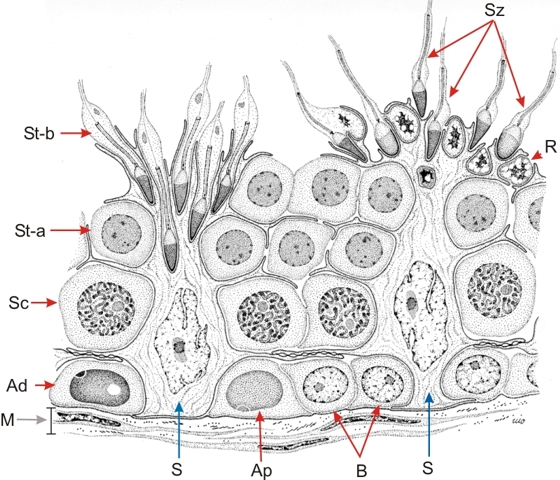
Drawing showing a small segment of a human seminiferous tubule.

Its seminiferous epithelium is composed of two main classes of cells: non-dividing somatic cells, or Sertoli cells (S), and germinal cells, that is, the spermatogonia (Ad, Ap and B) seen along the limiting membrane (M), the pachytene spermatocytes (Sc), the early round spermatids (St-a); the late elongated spermatids (St-b) and the spermatozoa (Sz).

Sertoli cell functions are the following:

  1. They are permanent supportive elements.
  2. They provide nutritive substances to the germinal cells.
  3. They secrete the luminal fluid.
  4. They are endocytic.
  5. They are phagocytic and eliminate residual bodies (R).
  6. They form a blood-testis barrier between the spermatogonia and the spermatocytes.


We'd very much appreciate receiving your comments and suggestions!We'd very much appreciate receiving your comments and suggestions!

Creative Commons Licence The text and images of this Histology Atlas, by Yves Clermont, Michael Lalli & Zsuzsanna Bencsath-Makkai, are licensed under a Creative Commons Attribution-Noncommercial-No Derivative Works 2.5 Canada Licence and cannot be modified without the written permission of the authors. Use of any text or images must carry an acknowledgement which includes a link to the original work.


HAweb/Zs B-M -> HAbase-generated on 2013-09-13