Contents Yves Clermont, Michael Lalli, Zsuzsanna Bencsath-Makkai
LIGHT-MICROSCOPIC HISTOLOGY ATLAS
     15. Male Reproductive System
  1     2     3     4     5     6     7     8     9     10     11     12     13     14     15     16     17     18     19     20     21     22     23     24     25  
  26     27     28     29     30     31     32     33     34     35     36     37     38     39     40     41     42     43     44  

    15.8   Human spermatogonia Version française
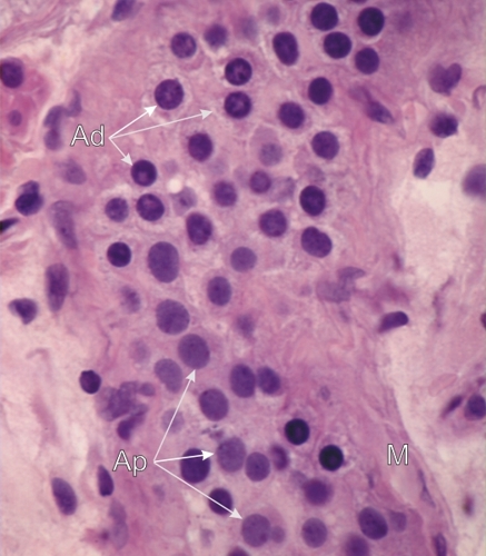
Tangential section through a human seminiferous tubule at the level of the basal layer of spermatogonia just above the limiting membrane (M).

The layer of spermatogonia is seen in face view. Among them the two main types of spermatogonia can be identified:

  • The dark type A spermatogonia or reserve stem cells (Ad) which rarely divide. Their intensely stained nuclei show a lightly stained central spherical cavity.
  • The pale type A spermatogonia or renewing stem cells (Ap) with their ovoid nuclei containing lightly stained homogeneous and finely granulated chromatin. These type Ap cells divide regularly by mitosis to produce new type Ap cells or type B spermatogonia.

Stain: H–­­E
Magnification: ×900


We'd very much appreciate receiving your comments and suggestions!We'd very much appreciate receiving your comments and suggestions!

Creative Commons Licence The text and images of this Histology Atlas, by Yves Clermont, Michael Lalli & Zsuzsanna Bencsath-Makkai, are licensed under a Creative Commons Attribution-Noncommercial-No Derivative Works 2.5 Canada Licence and cannot be modified without the written permission of the authors. Use of any text or images must carry an acknowledgement which includes a link to the original work.


HAweb/Zs B-M -> HAbase-generated on 2013-09-13